
Pregnancy leaves its mark on every body, yet most changes remain invisible. One hidden change that deserves attention is isthmocele which is a small pocket that can form inside the uterine wall after a cesarean section (C‑section).
On busy days you might not even think about the scar that helped deliver your baby, but if that scar heals unevenly it can create a niche that collects menstrual blood, sparks chronic pelvic discomfort, and even blocks future pregnancies.
With C‑section rates rising worldwide, more families now face questions about this condition than ever before. What does “uterine isthmocele” really mean? Why does it happen? When does it become a problem, and how can modern care restore comfort and fertility?
What Is an Isthmocele?
An isthmocele is a pouch‑like defect that forms in the lower part of the uterus, right where a previous C‑section incision was stitched closed.
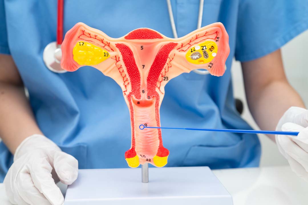
Think of the uterus as a muscular balloon. During a cesarean birth, surgeons create two cuts, one in the abdomen and another in the womb itself.
Ideally that uterine cut heals into a smooth, strong layer. Sometimes, though, the tissue sinks inward and leaves a small cave. Blood and fluid can pool inside, and over months or years the niche may widen.
Why the Lower Uterine Segment Matters
The defect almost always sits in the lower uterine segment (the “isthmus”). This area stretches thin at term, making it easier for surgeons to reach the baby quickly, but its limited blood supply can slow repair.
When healing lags or mechanical stress returns too soon, tissue edges fail to knit together, setting the stage for a visible gap on later imaging studies.
How Common Is Uterine Isthmocele?
Researchers once believed the problem was rare. Now, with better ultrasound techniques and rising cesarean rates, estimates sit between 24 percent and 70 percent among people with at least one C‑section.
Why the huge range? Diagnostic criteria differ, and many small niches never cause symptoms, so prevalence changes with the sensitivity of the scan. Still, even the lower estimate means roughly one in four post‑cesarean patients could carry a hidden scar pouch.
What Are the Main Isthmocele Causes?
Incomplete healing is the root, but several factors raise the chances of developing an isthmocele:
- Suture technique: One row of stitches (single‑layer closure) seals the incision faster but may leave weaker support than a double‑layer closure.
- Incision position: Very low cuts, often used when an emergency cesarean happens late in labor, sit right where the muscle is thinnest.
- Multiple surgeries: Each new C‑section reopens the same spot, increasing scar burden.
- Infections or sluggish healing: Diabetes, obesity, autoimmune disorders, or smoking can all slow tissue repair.
- Adhesions pulling on the scar: Internal scar bands may tug the edges apart over time.
We can talk to care providers about preventive strategies before a first or repeat cesarean by understanding these isthmocele causes
What Symptoms of Isthmocele Can You Notice?
Many niches remain silent, but the most common early clue is brown spotting for a few days after your normal period ends.
That dark flow is leftover blood draining from the pouch. Other signs include:
- Fresh bleeding between cycles or after intercourse.
- New or worsening period cramps.
- A sensation of pelvic pressure or fullness.
- Pain during sex.
- Mysterious vaginal discharge unrelated to infection.
If you’re trying to conceive, the niche can hinder sperm movement, trap embryos, or foster inflammation that blocks implantation. Recurrent miscarriage or infertility after a successful pregnancy may also hint at a hidden scar pocket.
How Can an Isthmocele Affect Fertility and Pregnancy Safety?
When menstrual blood stagnates in the pouch, inflammatory molecules build up. These chemicals can:
- Decrease sperm survival as they swim toward the tubes.
- Change the uterine environment so embryos fail to implant.
- Trigger immune reactions that harm early pregnancies.
Risks in Future Pregnancy
A thin scar segment makes rare yet serious events more likely:
- Scar ectopic pregnancy: The embryo implants inside the niche rather than the healthy lining.
- Placenta previa: The placenta covers the cervix.
- Placenta accreta: The placenta grows too deeply into the scar.
- Uterine rupture: In extreme cases, labor contractions can tear the weakened wall.
Proper diagnosis and isthmocele treatment before conception can dramatically reduce these dangers.
How Do Doctors Detect an Isthmocele? (Isthmocele Radiology)
Imaging is central to spotting the defect. Because a pouch looks most obvious right after the period (when it fills with residual blood), providers often schedule scans in the early follicular phase.
Transvaginal Ultrasound (TVUS or Isthmocele USG)
A bedside probe creates real‑time images. A triangular or semicircular niche appears in the lower uterine segment. Sonographers measure depth, width, and residual myometrium (the muscular layer) below the defect. A wall thinner than 3–5 mm deserves close attention.Saline Infusion Sonohysterography (SIS)
Saline expands the cavity like contrast dye, outlining even small niches. This test delivers more detail than a standard scan and helps distinguish between a true pouch and other anomalies, such as intrauterine adhesions.Hysteroscopy
A slim camera inserted through the cervix lets providers see the niche directly. If needed, they can treat minor defects during the same session.MRI
Reserved for complex cases, magnetic resonance imaging maps deeper pelvic anatomy and rules out scar pregnancies.Collectively, these tools fall under isthmocele radiology and guide personalized care.
Why Does Isthmocele USG Measurement Matter?
Isthmocele can be visualized and measured using ultrasound (USG). Measurements typically include the depth, width, length, and residual myometrial thickness at the defect site.
Ultrasound can also be used to classify isthmocele based on size, often using a system like:
- Grade 1: Less than 15 mm².
- Grade 2: 16 to 25 mm².
- Grade 3: Greater than 25 mm².
Numbers can affect decisions. A wall ≥ 5 mm suggests decent strength for future pregnancy; a thinner wall implies elevated risk. Depth and width also influence symptom severity as larger pockets trap more blood, leading to heavier post‑menstrual spotting.
What Are the Treatment Options for Isthmocele?
There are several treatment options available for isthmocele They are as follows:
Watchful Waiting and Hormonal Therapy
If bleeding is mild and fertility is not a priority, providers may prescribe low‑dose hormonal pills or a progesterone‑releasing intrauterine device. Hormones lighten periods, so less blood collects in the niche. Some small pouches shrink enough that surgery becomes unnecessary.Hysteroscopic Resection
Using a scope through the cervix, surgeons shave or cauterize the recess so blood can drain freely. This option suits people with a wall at least 5 mm thick and who either do not wish to conceive or have adequate myometrial thickness for safe pregnancy without full reconstruction.Laparoscopic Isthmocele Repair
For individuals who crave symptom relief and future fertility, laparoscopic isthmocele repair is the gold standard. Surgeons remove the entire pocket from the outside and rebuild the uterine wall in two or three muscular layers.Open Repair or Hysterectomy
Open surgery enters the discussion only for very large defects, extensive adhesions, or where laparoscopy is unavailable. Hysterectomy, the removal of the uterus, remains the last resort, considered when severe pain persists and childbearing is complete.What Happens During Laparoscopic Isthmocele Repair?
Step‑by‑step overview:
- You receive general anesthesia.
- Surgeons place tiny incisions in the abdomen for a camera and instruments.
- They identify the niche, remove scar tissue, and freshen the edges.
- The wall is reconstructed in layered stitches, restoring thickness.
- Instruments are withdrawn and the skin incisions closed.
Isthmocele Repair Recovery Time
Most patients stay one night or return home the same day. Light activity resumes in a week, full exercise by two weeks, and sexual intercourse once any spotting stops. Because the uterine muscle needs time to gain strength, providers recommend waiting about four months before trying to conceive.
How Can You Prepare for Isthmocele Surgery?
Some practical tips to prepare for isthmocele surgery:
- Stop smoking well in advance to improve oxygen delivery to healing tissue.
- Optimize weight and manage blood sugar if diabetic.
- Discuss all medications, especially blood thinners and hormonal therapies.
- Arrange help at home for the first few days, particularly if you have small children.
- Plan for pelvic rest — no tampons, sex, or swimming until cleared.
Preventing a New Isthmocele During Future Births
If you need another cesarean, discuss preventive measures with your surgical team:
- Request a double‑layer closure if clinically safe.
- Ask whether the incision can be placed slightly higher in the muscular portion.
- Allow adequate spacing between pregnancies to support full scar maturation (ideally at least 18 months).
- Maintain healthy lifestyle habits that promote collagen formation and tissue repair.
Frequently Asked Questions
Yes. Post-menstrual brown discharge is the classic sign because old blood dribbles out of the niche days after the main flow ends.
No. Many people conceive naturally despite a niche, but the risk of implantation failure or miscarriage rises as pocket size grows.
Hormones can reduce bleeding and minor discomfort but rarely close the pouch entirely. They act as symptom control, not structural repair.
Most providers still recommend a planned cesarean for subsequent births to protect the reconstructed wall, though individual advice varies.
You may feel mild cramping similar to menstrual pain for a day or two. Over‑the‑counter pain relievers usually suffice.
That thickness poses a higher rupture risk in pregnancy. Laparoscopic repair is often advised before attempting to conceive.
Implantation can fail and miscarriage risk is higher. Repairing the defect before IVF is usually recommended.
Conclusion
An isthmocele may be tiny, yet its reach can be profound like disrupting cycles, causing pain, and standing between you and future plans for a family. The good news is that modern imaging pinpoints the defect promptly, and minimally invasive techniques rebuild the uterine wall with excellent success rates.
If you notice lingering spotting, pelvic pressure, or fertility hurdles after a C‑section, trust your instincts and seek evaluation. A simple ultrasound could reveal a pocket that is both diagnosable and fixable.
From hormonal tweaks to sophisticated laparoscopic reconstruction, today’s options put you back in control of your health journey. With informed choices and timely care, you can move past the scar, reclaim comfort, and look forward with confidence to safe pregnancies or simply symptom‑free living.

About Us
AKsigen IVF is a premier center for advanced fertility treatments, with renowned fertility experts on our team. Specializing in IVF, ICSI, egg freezing, and other cutting-edge reproductive technologies, AKsigen IVF is committed to helping couples achieve their dream of parenthood. With personalized care and a patient-first approach, AKsigen IVF provides comprehensive fertility solutions under one roof.



 +91 86556 78747
 info@aksigenivf.com


